
Volunteer by tracing, labeling, and reviewing medical images for real-world research.
Train on ISIC 2018 lesion images, annotate in your browser, and earn volunteer credit for eligible submissions.
How volunteering works
You’ll learn lesion boundary segmentation, then complete pilot tasks scored against ISIC reference masks.
Create your account- 01
Training
Review examples and instructions for a specific task, such as tracing a border or marking a visible structure.
- 02
Image annotation
Label the requested visual features in medical images. Volunteers do not diagnose patients or make medical decisions.
- 03
Volunteer credit
After training, verified students annotate ISIC images. Each unique submission with a Dice score greater than 0.9 earns 2 volunteer minutes.
Task-specific trainingExamples and instructions before starting
Researcher reviewFor submitted annotations
Volunteer hoursRecorded for accepted work
Example image tasks
Tasks depend on the research project. These examples show image types and features volunteers may annotate.
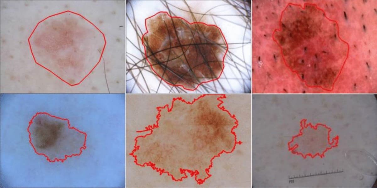
Lesion boundary tracing
Trace the visible edge of a skin lesion for a researcher to review.
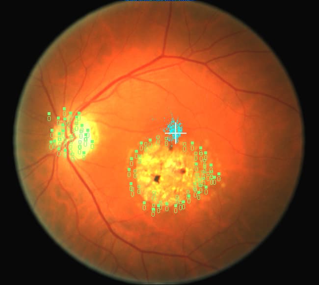
Fundus landmark marking
Mark vessels, the optic disc, or other specified features in fundus photos.
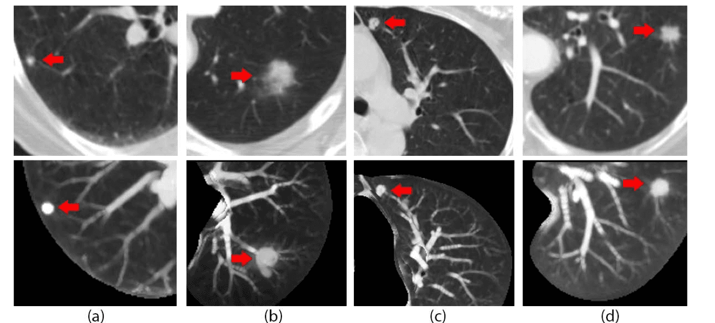
Cross-sectional region review
Flag specified regions in CT slices for a researcher to inspect.
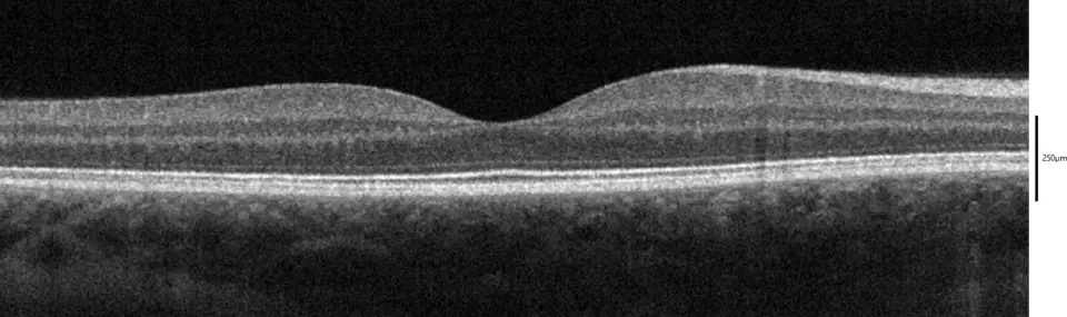
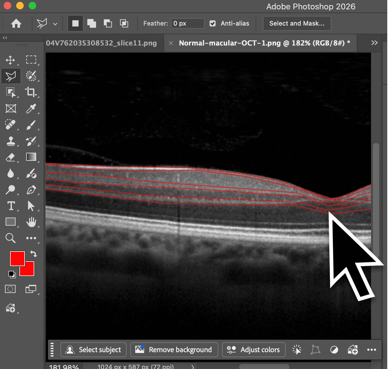
Layer segmentation support
Trace visible retinal layer boundaries in OCT scans.
Registration
Create an account, complete the training, and get ready for your first annotation after enrollment approval.
Join the annotation pilot Information for research labs