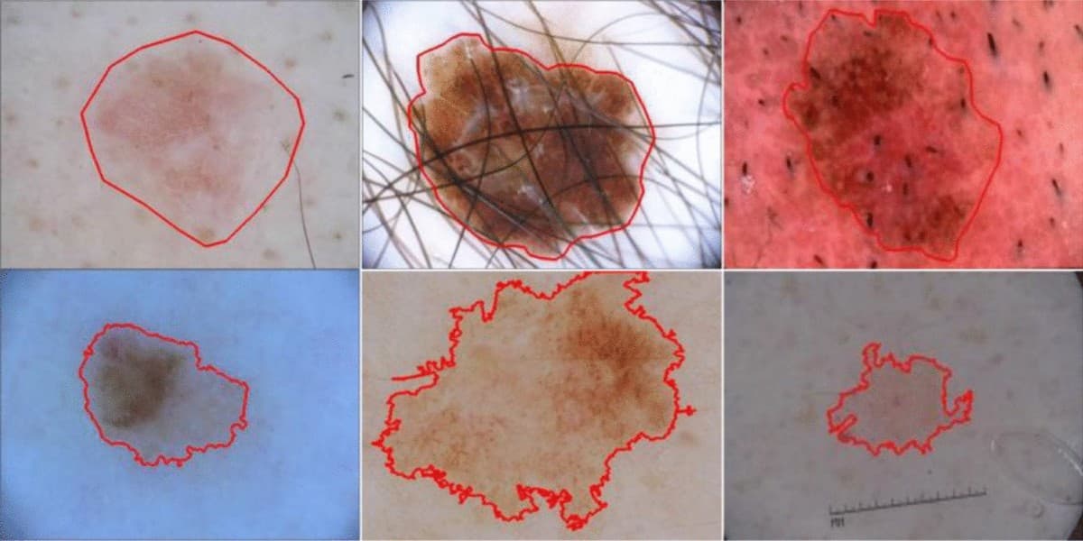
Dermoscopic skin lesion
Untrained contributors achieved good-to-excellent agreement with experts when identifying six visible dermoscopic features (κ = 0.71–0.84).
Kentley et al., 2023
 Back to site
AnnotationBridge prepares student volunteers for defined image annotation tasks. Research teams provide the task instructions and review submitted work.
Volunteers can trace visible structures and label specified features in de-identified medical images.
Volunteers do not make clinical decisions. Researchers approve the annotations, and students receive volunteer hours for accepted work.
These studies examine how non-experts perform defined image annotation tasks with training or expert comparison.
Untrained contributors achieved good-to-excellent agreement with experts when identifying six visible dermoscopic features (κ = 0.71–0.84).
Kentley et al., 2023
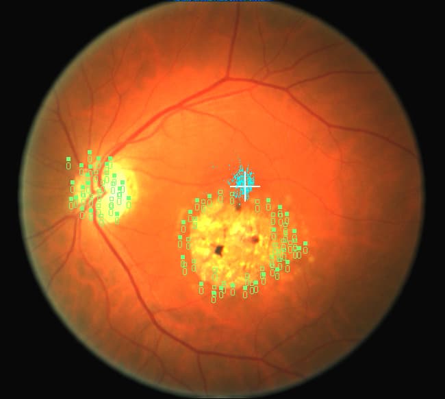
Ophthalmology-naive annotators produced abnormality labels comparable to experts; combined classifications reached an AUC of 0.93.
Mitry et al., 2016
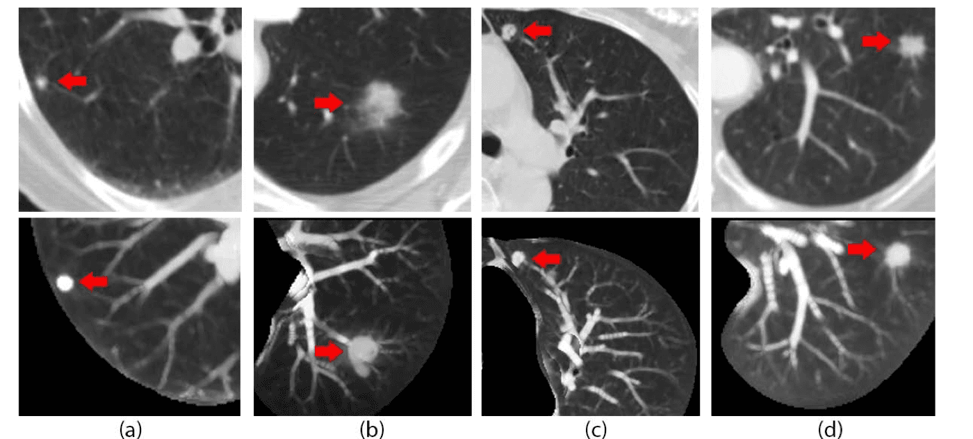
After a brief tutorial, non-experts detected lung nodules with over 90% sensitivity, and 96% for nodules larger than 4 mm.
Boorboor et al., 2018